
CO₂ Nanobubbles: Nanobubble Technology for Health Science
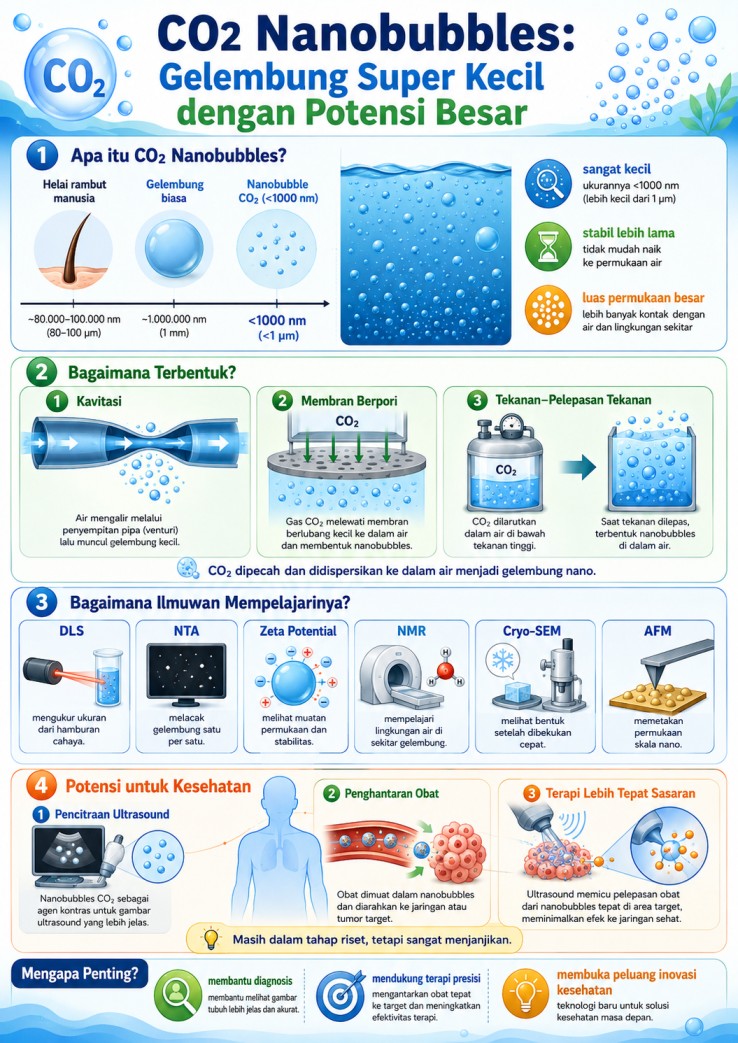
Nanobubbles are gas bubbles at the nanoscale, generally smaller than 1,000 nanometers. Their size is extremely small, but this is what gives them unique properties. These bubbles have a large surface area, low buoyancy, high internal pressure, and can remain in liquid longer than ordinary bubbles. One type that is currently being widely studied is CO₂ nanobubbles, which are nano-sized bubbles filled with carbon dioxide and dispersed in water. This technology was initially discussed mainly in the context of energy, the environment, and the food industry. However, in recent years, nanobubbles have also begun to attract attention in health and biomedical science.
How Are CO₂ Nanobubbles Formed?
To produce CO₂ nanobubbles, carbon dioxide gas cannot simply be introduced into water. The gas must be dispersed into very small, stable bubbles that are evenly distributed throughout the liquid. This process is influenced by thermodynamic and kinetic factors, meaning that its success depends not only on the equipment used, but also on pressure, temperature, pH, salt concentration, the type of liquid, and additives in the solution.One of the main methods for forming CO₂ nanobubbles is cavitation, a process in which liquid undergoes rapid pressure changes that lead to the formation of small bubbles. Cavitation can be carried out through hydrodynamic cavitation, for example by flowing liquid through a venturi tube or perforated plate, and through acoustic cavitation, which uses ultrasonic waves. This method is widely used because it is relatively simple, has potential for large-scale application, and can produce very small bubbles.
Another method is membrane dispersion, in which CO₂ gas is passed through a porous membrane into a liquid. When the gas passes through the membrane pores and meets the liquid phase, it breaks into fine bubbles. The advantage of this method is that bubble size can be controlled more easily because it is influenced by membrane pore size, gas pressure, liquid flow rate, and the properties of the solution. For this reason, membrane dispersion is promising for applications that require more uniform bubble sizes. CO₂ nanobubbles can also be produced using the pressurization–depressurization method. In this process, CO₂ is first dissolved in water or another solution under high pressure. The pressure is then rapidly reduced, causing the dissolved gas to form small bubbles. Commercial nanobubble generators are also now available, combining the principles of cavitation, pressure, liquid flow, or membrane systems to produce nanobubbles more practically and consistently.
In general, each method has its own advantages and limitations. Cavitation is strong in terms of simplicity and scalability. Membrane dispersion is superior for size control. Pressurization–depressurization is useful because it takes advantage of gas solubility under pressure, while commercial generators offer a more practical approach. For sensitive applications such as health, the formation of CO₂ nanobubbles must be carefully controlled so that their size, concentration, stability, purity, and solution composition remain consistent.
How Do Scientists Prove That Nanobubbles Exist?
CO₂ nanobubbles attract researchers’ attention because they can remain in liquid longer than ordinary bubbles. In theory, very small bubbles should quickly dissolve or disappear. However, nanobubbles have several properties that make them more stable. Their extremely small size gives them low buoyancy, so they do not easily rise to the surface. In addition, the surface of nanobubbles often carries an electric charge. This charge causes the bubbles to repel each other, making it more difficult for them to merge into larger bubbles. Their stability is also affected by liquid conditions such as pH, salt concentration, surfactants, temperature, and other dissolved substances. In CO₂ nanobubbles, stability is also related to the interaction between CO₂ and water. When CO₂ is present in water, part of the gas can react and form compounds such as carbonic acid, bicarbonate, and carbonate. This process can affect the pH of the solution as well as the surface properties of the bubbles. If the pH and solution composition are supportive, nanobubbles can become more stable. On the other hand, unsuitable solution conditions can cause bubbles to grow, merge, or disappear more easily. This is why understanding water chemistry is important in developing CO₂ nanobubbles, especially for sensitive applications such as health.
Because their size is at the nanoscale, CO₂ nanobubbles cannot be observed directly with the naked eye. Scientists need special characterization techniques to prove their presence, measure their size, count their number, and understand their behavior in liquid. One commonly used technique is Dynamic Light Scattering, or DLS. This technique uses laser light directed at a liquid sample. When the light hits nanobubbles, it is scattered. From this scattering pattern, researchers can estimate the average bubble size. DLS is useful for observing nanobubble size distribution, but its results must be interpreted carefully because the instrument can also detect tiny impurity particles in the liquid. Another commonly used technique is Nanoparticle Tracking Analysis, or NTA. Unlike DLS, which reads the overall scattering pattern, NTA tracks the movement of individual bubbles using a camera. From the random movement of nanobubbles in the liquid, the instrument can calculate the size and number of bubbles in the sample. This information is important because researchers need to know not only how large the bubbles are, but also how many bubbles are formed and whether their number remains stable over time. For this reason, NTA is often used to monitor nanobubble concentration and changes in size during storage or after treatment.
In addition to size and number, scientists also need to understand the surface charge of nanobubbles. This is measured using zeta potential. The zeta potential value indicates whether bubbles tend to repel each other or merge. If the surface charge is strong enough, the bubbles are less likely to combine, and the system becomes more stable. However, if the value is close to neutral, nanobubbles are more likely to merge, grow larger, and disappear. In CO₂ nanobubbles, zeta potential is strongly influenced by pH, salt concentration, surfactant type, and water composition. This technique is important for designing solutions that can keep nanobubbles stable for longer periods. To understand the molecular environment of water around nanobubbles, researchers can use Nuclear Magnetic Resonance, or NMR. In nanobubble research, NMR helps scientists study how water molecules move and interact around the bubble surface. This information is important because the nanobubble surface is the main area where CO₂, water, and ions in the solution interact. NMR not only provides information about the presence of bubbles, but also helps explain how nanobubbles can affect the structure and mobility of water around them.
Another technique used to observe the shape of nanobubbles is cryo-scanning electron microscopy, or cryo-SEM. In this technique, the sample is rapidly frozen so that the structures inside the liquid can be preserved before being observed using an electron microscope. Cryo-SEM can provide visual information about the shape and morphology of nanobubbles. However, the results must be analyzed carefully because the freezing process can affect the position or shape of the bubbles. In addition, cryo-SEM shows physical structure, but it does not always confirm the gas composition inside the bubble. Meanwhile, Atomic Force Microscopy, or AFM, is used to map surfaces at the nanoscale. In nanobubble research, AFM can help observe nanobubbles located on solid surfaces or at the interface between a liquid and a surface. This technique can provide information about bubble shape, height, stiffness, and how nanobubbles attach to or change on a surface. AFM is useful for understanding nanobubble behavior in contact areas, especially when nanobubbles are used in systems involving biological surfaces, medical materials, or membranes.
Overall, no single technique can fully explain all the properties of CO₂ nanobubbles. DLS helps measure average size, NTA counts the size and number of bubbles one by one, zeta potential explains stability based on surface charge, NMR observes water molecule interactions, cryo-SEM provides shape information after freezing, and AFM maps surfaces at the nanoscale. By combining these techniques, researchers can gain a more complete understanding of how CO₂ nanobubbles form, persist, change, and potentially be used in health applications.
The Potential of CO₂ Nanobubbles for Health
In the health field, CO₂ nanobubbles are being studied because of their very small size and their ability to interact with fluids and biological tissues. Nanobubbles have potential uses in several biomedical applications, such as supporting ultrasound imaging, carrying drugs to specific areas, and enabling therapies that can be activated by stimuli such as ultrasound waves. Although the potential is significant, much of this technology is still at the research stage. Its use in humans still requires strict testing for safety, effectiveness, dosage, stability, and regulatory compliance. One of the main opportunities for CO₂ nanobubbles is as contrast agents for ultrasound imaging. In ultrasound examinations, sound waves are used to produce images of organs or tissues. Gas bubbles can help reflect sound waves, potentially making the resulting images clearer. Because nanobubbles are smaller than microbubbles, they may theoretically move more easily through small blood vessels and reach areas that larger bubbles may not be able to access.
CO₂ nanobubbles are also interesting for development as a more targeted drug delivery system. Drugs can be designed to attach to the surface of nanobubbles or be carried together with them. When the nanobubbles reach a target area, such as tumor tissue, ultrasound waves can be used to help release the drug at the desired location. This approach may improve therapeutic effectiveness and reduce drug exposure to healthy tissue, but the design must be handled very carefully because CO₂ can affect pH and the chemical conditions around tissues. In addition, nanobubbles can respond to ultrasound waves through a process called cavitation, which refers to changes in bubble behavior when exposed to sound waves. In biomedical research, this process is being studied to help improve drug penetration, temporarily open cell membranes, or support more targeted therapy. However, cavitation effects must be precisely controlled to avoid damaging healthy tissue.
Nanobubbles are also being studied as gas carriers and as tools for diagnostics and cancer therapy. Some tumor tissues have blood vessel structures that allow small particles to enter and remain in the area more easily. This opens the possibility of using nanobubbles to carry imaging agents or anticancer drugs. With the help of ultrasound, nanobubbles may help clarify the target location while supporting more localized drug release. The biggest challenge in using CO₂ nanobubbles in health applications is ensuring that the technology is safe, stable, and can be produced consistently. Blood, proteins, salts, cells, and body fluids can alter the properties of nanobubbles. In addition, because CO₂ can affect pH, its concentration and release rate must be carefully controlled. Therefore, accurate characterization using techniques such as DLS, NTA, zeta potential, NMR, cryo-SEM, and AFM is needed, along with sterile and uniform production from one batch to the next. If these challenges can be addressed, CO₂ nanobubbles may become part of future health technologies, especially for medical imaging, drug delivery, and ultrasound-assisted therapy.
Source:
Kumar, A., Paker, D. M., Tamerler, C., Mountziaris, T. J., & Dindoruk, B. (2025). Recent advancements in aqueous CO2 nanobubbles. Energy & Fuels, 39(38), 18194–18217. https://doi.org/10.1021/acs.energyfuels.5c01218